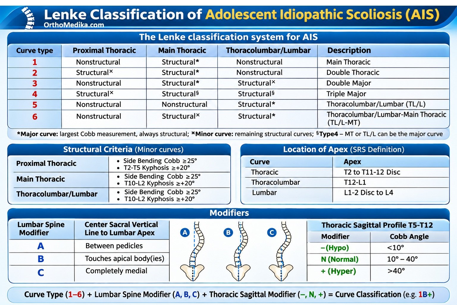
The Lenke classification for adolescent idiopathic scoliosis (AIS) is a three-part code: curve type (1–6) + lumbar modifier (A/B/C) + sagittal thoracic modifier (−/N/+). For example, Lenke 1A− means a single main thoracic structural curve, a relatively centered lumbar curve, and reduced thoracic kyphosis.
What you need first
Use standing full-spine radiographs:
- PA/AP radiograph: measures Cobb angles and determines the lumbar modifier.
- Left and right side-bending radiographs: determine whether minor curves are structural.
- Lateral radiograph: measures thoracic kyphosis for the sagittal modifier.
Divide the spine into three regions:
| Region | Curve apex |
| Proximal thoracic (PT) | T3, T4, or T5 |
| Main thoracic (MT) | T6 through the T11–T12 disc |
| Thoracolumbar/lumbar (TL/L) | T12–L1 disc through L4 |
The apex is the most rotated and most deviated vertebra or disc in a curve.
Step 1: Measure the curves
Measure the Cobb angle for each of the three regions:
- Identify the most tilted vertebra above the curve—the upper end vertebra.
- Identify the most tilted vertebra below the curve—the lower end vertebra.
- Draw lines along the upper endplate of the upper end vertebra and lower endplate of the lower end vertebra.
- Measure the angle between perpendiculars to those lines; this is the Cobb angle.
The curve with the largest Cobb angle is the major curve. It is structural by definition. The other curves are minor curves until proven structural.
Step 2: Decide which curves are structural
A minor curve is structural if it remains 25° or more on side-bending films. Conversely, a minor curve that corrects to less than 25° is nonstructural.pmc.ncbi.nlm.nih+1
There are two additional sagittal rules:
- A proximal thoracic curve is structural if T2–T5 kyphosis is 20° or more.
- A main thoracic or thoracolumbar/lumbar curve is structural if T10–L2 kyphosis is 20° or more.
So, for each region, record:
- Cobb angle on standing PA film
- Cobb angle on bending films
- Whether it is structural or nonstructural
- Whether it is the major curve
Step 3: Assign the curve type
Use the pattern of structural curves and identify the major curve.
| Lenke type | Pattern | Major curve | Structural curves |
| 1 | Main thoracic | MT | MT only |
| 2 | Double thoracic | MT | PT + MT |
| 3 | Double major | MT | MT + TL/L |
| 4 | Triple major | MT | PT + MT + TL/L |
| 5 | Thoracolumbar/lumbar | TL/L | TL/L only |
| 6 | TL/L–main thoracic | TL/L | TL/L + MT |
Important distinction: in Type 3, the MT curve is larger; in Type 6, the TL/L curve is larger by at least 5°, but the MT curve is still structural.
A simple way to remember:
- Type 1: one thoracic curve.
- Type 2: two thoracic curves.
- Type 3: thoracic + lumbar, thoracic larger.
- Type 4: three structural curves.
- Type 5: one lumbar/TL curve.
- Type 6: thoracic + lumbar, lumbar larger.
Step 4: Assign the lumbar modifier
Draw the center sacral vertical line (CSVL): a vertical line upward from the center of the sacrum on the PA radiograph. Then look at its relationship to the apical vertebra of the lumbar or thoracolumbar curve.
| Modifier | CSVL position at lumbar apex | Meaning |
| A | Passes between the pedicles | Lumbar curve is well centered |
| B | Touches the apical pedicle or vertebral body | Intermediate lumbar shift |
| C | Lies completely medial to the apical vertebral body/pedicle | Lumbar curve is substantially shifted |
In practice: A = centered, B = borderline, C = decompensated/lumbar shift.
Step 5: Assign the sagittal thoracic modifier
On the standing lateral radiograph, measure thoracic kyphosis from the upper endplate of T5 to the lower endplate of T12.
| Modifier | T5–T12 kyphosis | Description |
| − | Less than 10° | Hypokyphosis |
| N | 10° to 40° | Normal kyphosis |
| + | More than 40° | Hyperkyphosis |
This modifier is important because it describes the sagittal profile, not just the sideways curve.
Step 6: Write the final classification
Combine the three parts:
Examples:
- Lenke 1A−: single structural main thoracic curve; CSVL between lumbar pedicles; T5–T12 kyphosis <10°.
- Lenke 1BN: single main thoracic curve; CSVL touches the lumbar apex; normal thoracic kyphosis.
- Lenke 3C+: double major curve with MT as major; substantial lumbar shift; thoracic hyperkyphosis.
- Lenke 5C−: single structural TL/L curve; lumbar shift; hypokyphosis.
Worked example
Suppose the PA film shows:
- PT curve: 20°
- MT curve: 50°
- TL/L curve: 35°
The MT curve is major because 50° is largest.
On bending films:
- PT corrects to 18° → nonstructural
- TL/L corrects to 28° → structural
Therefore, structural curves are MT + TL/L, and the MT is major → Lenke Type 3.
Next, the CSVL passes completely medial to the lumbar apical vertebra → lumbar modifier C.
The lateral film shows T5–T12 kyphosis of 18° → N.
Final classification: Lenke 3CN
REFERENCES:
AO Surgery References : https://surgeryreference.aofoundation.org/spine/deformities/adolescent-idiopathic-scoliosis/further-reading/lenke-classification#table-summary
Orthobullets : https://www.orthobullets.com/spine/2076/lenke-classification-of-ais

 is a three-part code: curve type (1–6) + lumbar modifier (A/B/C) + sagittal thoracic modifier (−/N/+).)