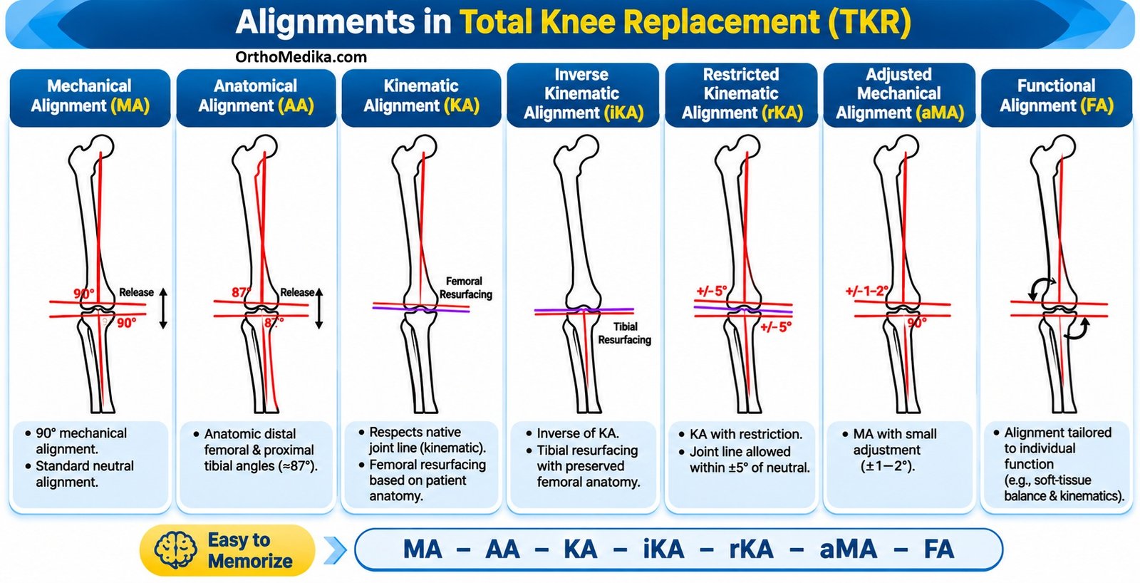
TKA alignment means positioning the femoral, tibial, and sometimes patellar components so that the reconstructed knee is stable, mobile, and durable. It is not only about making the leg straight; it also includes the coronal plane (varus–valgus), sagittal plane (flexion–extension), and axial/rotational plane (femoral and tibial rotation).
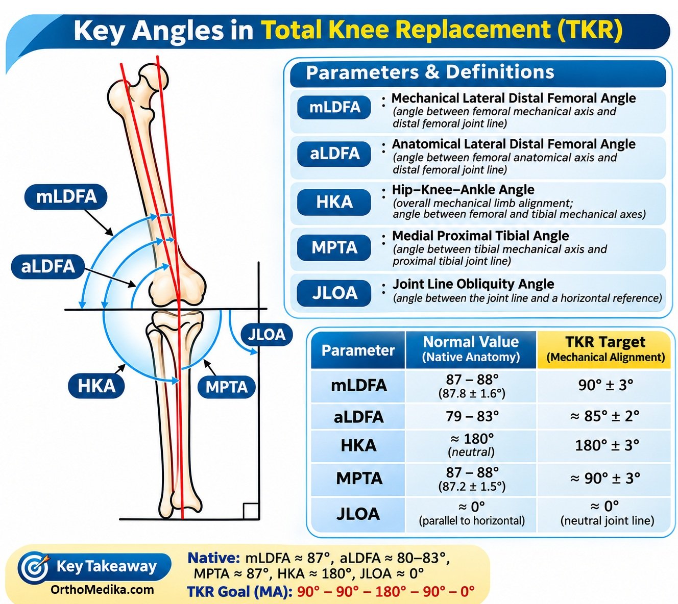
The most important coronal measurement is the hip–knee–ankle (HKA) angle:
- 180° = neutral mechanical axis
- <180° = varus alignment
- >180° = valgus alignment
The Matassi et al. review groups alignment strategies into three families: systematic alignment (mechanical and anatomic), patient-specific alignment (kinematic), and hybrid/personalized alignment (restricted KA, inverse KA, adjusted mechanical alignment, and functional alignment).
Why alignment matters
The classical rationale for neutral alignment is biomechanical: a straight limb theoretically distributes load evenly between the medial and lateral tibial compartments, reducing asymmetric polyethylene wear, component loosening, and revision risk.pmc.ncbi.nlm.nih+1
However, forcing every patient into neutral alignment may conflict with that patient’s native anatomy. In a patient with constitutional varus, correcting the limb to 180° can require ligament release, alter joint-line obliquity, change femoral flexion kinematics, and affect patellar tracking.
Therefore, modern TKA alignment has shifted from asking, “Is the leg perfectly straight?” to asking, “Is the knee stable, balanced, naturally functioning, and positioned within a safe range?”
Key anatomical references
| Term | Meaning | Why it matters |
| HKA angle | Angle between femoral mechanical axis and tibial mechanical axis | Describes overall limb alignment; 180° is neutral |
| mLDFA | Distal lateral femoral angle; angle between femoral mechanical axis and distal femoral joint line | Describes femoral component/joint-line orientation |
| MPTA | Medial proximal tibial angle; angle between tibial mechanical axis and proximal tibial joint line | Describes tibial component/joint-line orientation |
| Joint-line obliquity (JLO) | Obliquity of the knee joint line relative to the floor | Affects ligament tension and knee kinematics; KA tends to preserve it more than MA |
| Flexion–extension gap | Space available for the implant with knee flexed and extended | Balanced gaps are essential for stability and motion |
| Safe zone | Acceptable range for HKA and component angles | Limits extreme varus/valgus while allowing personalization |
A. Mechanical alignment (MA)
Principle
Mechanical alignment aims to reconstruct every knee to a neutral limb axis: HKA 180°, with the femoral component perpendicular to the femoral mechanical axis and the tibial component perpendicular to the tibial mechanical axis. The tibial cut is conventionally 90° to the tibial mechanical axis.
Surgical logic
- Establish the mechanical axes of femur and tibia.
- Make standardized bone cuts to place both components perpendicular to their respective mechanical axes.
- Achieve a neutral HKA.
- Use soft-tissue releases or balancing techniques to create equal medial and lateral gaps.
Main advantage
MA has the longest clinical experience and strongest long-term survivorship data. Its theoretical benefit is symmetric load distribution across the implant, which may reduce wear and aseptic loosening.
Main limitation
MA is a “one-size-fits-all” target. In patients with constitutional varus or valgus, it may require more soft-tissue release and can create a less natural joint line or alter native knee kinematics.
Example: In a patient with 8° constitutional varus, MA would correct the limb to 0° HKA. This may require medial soft-tissue release and may produce a joint line that differs from the patient’s pre-arthritic anatomy.
Anatomic alignment (AA)
Principle
Anatomic alignment is an older personalized concept that attempts to reproduce the normal anatomic orientation of the knee: approximately 6–7° femoral valgus and approximately 3° tibial varus, while still maintaining an overall neutral HKA of 180°.pmc.ncbi.nlm.nih+1
Surgical logic
- The femoral component is placed in about 3° valgus relative to the femoral mechanical axis.
- The tibial component is placed in about 3° varus relative to the tibial mechanical axis.
- The combined limb alignment remains neutral.
Difference from MA
MA makes both femoral and tibial cuts perpendicular to their mechanical axes. AA preserves the natural obliquity of each bone’s joint surface, but still targets neutral overall limb alignment.
Main limitation
AA is technically demanding because small errors in femoral or tibial cuts can compound and produce limb malalignment. It has largely been superseded by KA and hybrid techniques.
B. Kinematic alignment (KA)
Principle
Kinematic alignment seeks to resurface the patient’s pre-arthritic knee anatomy rather than impose a standard neutral alignment. It preserves the native limb alignment, joint-line obliquity, and soft-tissue envelope.
KA is based on the idea that the non-arthritic knee is already balanced. Therefore, if the surgeon restores the pre-arthritic anatomy and corrects only cartilage/bone loss caused by arthritis, the soft tissues should remain balanced without extensive release.
Surgical logic
- Identify the patient’s pre-arthritic alignment and joint-line orientation.
- Resurface the femur to preserve the native distal and posterior femoral joint lines.
- Restore the native tibial joint line, often accepting mild tibial varus if it matches the patient’s anatomy.
- Preserve the three rotational axes of the knee.
- Avoid routine ligament release unless instability persists.
Difference from MA
| Feature | Mechanical alignment | Kinematic alignment |
| Primary target | Neutral HKA 180° | Patient’s pre-arthritic anatomy |
| Femoral component | Perpendicular to femoral mechanical axis | Restores native femoral joint line and kinematics |
| Tibial component | 90° to tibial mechanical axis | Restores native tibial joint-line obliquity |
| Soft tissue | Often requires release | Usually preserves soft-tissue envelope |
| Joint line | Often horizontal/neutral | More physiologic/native obliquity |
| Main risk | Instability, altered kinematics, need for release | Excessive tibial varus or alignment outliers if unrestricted |
Evidence
KA produces radiographically non-neutral alignment compared with MA: systematic reviews report approximately 1.9° more tibial varus and 1.6° more femoral valgus. Clinical outcomes are generally comparable to MA, with some studies showing better pain, flexion, function, or patient satisfaction, but evidence remains mixed and long-term superiority is not established.
C. Restricted kinematic alignment (rKA)
Principle
rKA is KA with guardrails. It preserves the patient’s anatomy, but only within predefined limits to avoid dangerous outliers.
A commonly described rKA framework uses:
- HKA within ±3° of neutral, i.e., 177°–183°
- mLDFA and MPTA within ±5° of target
- Tibial cut restricted to 84° varus to 92° valgus relative to the tibial mechanical axis.
Difference from unrestricted KA
Unrestricted KA may follow the patient’s native anatomy even if it results in marked varus. rKA allows personalization but prevents excessive tibial varus or overall limb varus/valgus that could threaten implant fixation.
Example: If a patient has 10° constitutional tibial varus, unrestricted KA might reproduce it, whereas rKA would limit the tibial correction to the safe zone and may accept partial correction or use soft-tissue balancing.
D. Inverse kinematic alignment (iKA)
Principle
iKA starts with the tibia rather than the femur. It uses a “true tibial resurfacing” approach, preserving the native tibial joint line within a restricted range, then adjusts the femoral component to balance the knee.
Surgical logic
- Restore the tibial joint line first.
- Limit tibial resection to a safe range, commonly 84° varus to 92° valgus.
- Adjust the femur to create balanced flexion and extension gaps.
- Maintain overall limb alignment within a safe zone, often described as approximately 174°–183° HKA.
Difference from conventional KA
Conventional KA is often described as “femur-first” or “true femoral resurfacing,” preserving the femoral joint line. iKA is “tibia-first,” preserving the tibial joint line and then balancing the femur.
Main limitation
iKA is a newer concept with limited long-term survivorship data.
E. Adjusted mechanical alignment (aMA)
Principle
Adjusted mechanical alignment is a hybrid between MA and KA. The tibial component remains perpendicular to the tibial mechanical axis, but the femoral component is adjusted to preserve some of the patient’s constitutional femoral anatomy and reduce the need for release.
Surgical logic
- Keep the tibial cut at 90° to the tibial mechanical axis.
- Modify the femoral cut to respect the patient’s constitutional deformity.
- Aim for slight undercorrection rather than forcing full neutral alignment.
Difference from MA and KA
| Feature | MA | aMA | KA |
| Tibia | 90° to mechanical axis | 90° to mechanical axis | Native/pre-arthritic orientation |
| Femur | Perpendicular to mechanical axis | Adjusted to patient anatomy | Native/pre-arthritic orientation |
| Soft tissue | May need release | Less release than MA in selected cases | Minimal release |
| Overall goal | Neutral limb | Slight undercorrection within safe zone | Restore native alignment |
F. Functional alignment (FA)
Principle
Functional alignment is a gap-balancing, soft-tissue-sparing personalized technique. Its priority is not a fixed radiographic target but a stable, balanced knee with minimal soft-tissue release.
FA tries to recreate the patient’s pre-arthritic condition by first creating a rectangular extension gap, then balancing the flexion gap through fine adjustments of femoral and tibial component position.
Surgical logic
- Plan the tibial and femoral resections.
- Create a rectangular extension gap at 0°.
- Balance the flexion gap.
- Fine-tune femoral and tibial component position to equalize medial and lateral gaps.
- Avoid soft-tissue release whenever possible.
Difference from KA
KA is primarily anatomy-driven: it restores the patient’s pre-arthritic bony anatomy and assumes the soft tissues will balance. FA is primarily soft-tissue/gap-driven: it adjusts component position to achieve balance while staying within safe alignment boundaries.
In one robotic-assisted study, FA achieved balanced flexion–extension gaps in 96.5% of knees, compared with 66.4% for KA and 54.7% for MA. This supports FA’s soft-tissue-balancing rationale, though it does not establish superior long-term implant survival.
Practical comparison
| Technique | Main target | Tibial cut | Femoral cut | Soft tissue | Main advantage | Main concern |
| MA | HKA 180° | 90° | Perpendicular to femoral mechanical axis | Release as needed | Proven long-term approach | More release; may alter native kinematics |
| AA | Neutral HKA with anatomic femur/tibia | About 87° | About 3° valgus | Variable | More natural component orientation | Technical difficulty; largely historical |
| KA | Pre-arthritic anatomy | Native orientation | Native orientation | Usually preserved | Natural kinematics, less release | Possible tibial varus/outliers |
| rKA | Native anatomy within safe zone | 84°–92° | Native within limits | Usually preserved | Personalization with safety limits | Less long-term evidence |
| iKA | Tibial joint line first | 84°–92° | Adjusted for balance | Usually preserved | Tibia-first logic | Newer, limited evidence |
| aMA | Tibia neutral, femur personalized | 90° | Adjusted | Less release than MA | Compromise between MA and KA | No proven superiority |
| FA | Balanced gaps, minimal release | According to extension gap, ±3° | Adjusted for balance | Minimal release | High rate of gap balance | Needs planning/navigation; long-term data limited |
What “safe zone” means
A safe zone is an accepted range of alignment that avoids extreme deviations while allowing individualized positioning. The Matassi review describes hybrid methods as aiming for an HKA safe zone of 177°–183°.
Other commonly cited limits include:
- HKA: 177°–183°, or ±3° from neutral
- mLDFA and MPTA: within ±5° of target
- Tibial component: avoid excessive varus, often limited to 84°–92° depending on technique
- Joint-line obliquity: preserve physiologic obliquity without creating instability
The purpose is to avoid the two extremes: forcing all knees to 180° when their anatomy does not permit it, and reproducing severe constitutional deformity that may overload one compartment.
Current evidence and consensus
There is still no definitive consensus that one alignment philosophy is superior for all patients. MA remains the traditional standard with the most long-term survivorship data, while KA, rKA, iKA, aMA, and FA are increasingly used to preserve soft tissues and improve gap balance.
Recent systematic reviews suggest KA may offer modest advantages in pain, flexion, and function in some analyses, but other reviews find no meaningful difference from MA. Complication rates are generally similar.