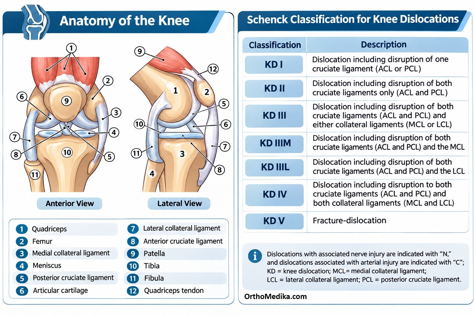
The Modified Schenck classification is the most widely used anatomic system for traumatic knee dislocations. It describes which major stabilizers are injured—ACL, PCL, MCL, and LCL/posterolateral corner (PLC)—and adds a fracture-dislocation category, making it useful for communication, surgical planning, and estimating neurovascular risk.
Basic knee anatomy
The knee is primarily a modified hinge joint with three articulations: tibiofemoral (femur–tibia), patellofemoral (patella–femur), and proximal tibiofibular (tibia–fibula). A true knee dislocation refers to complete loss of tibiofemoral congruity.
The four major ligament groups assessed in the Schenck system are:
| Structure | Attachments / location | Main function |
| ACL | Runs from the anterior tibial intercondylar area to the posteromedial surface of the lateral femoral condyle. | Prevents the tibia from translating anteriorly under the femur; limits hyperextension and rotational instability. |
| PCL | Runs from the posterior tibial intercondylar region to the anterolateral surface of the medial femoral condyle. | Prevents posterior tibial translation; the stronger cruciate ligament and important in flexed-weight-bearing positions. pmc. |
| MCL / posteromedial corner | Medial side of knee; superficial MCL, deep MCL, posterior oblique ligament, posteromedial capsule, and semimembranosus expansions. | Resists valgus force (a blow to the lateral knee that opens the medial side) and controls medial rotation. |
| LCL / posterolateral corner | Lateral side; fibular collateral ligament, popliteus tendon, popliteofibular ligament, biceps femoris, and posterolateral capsule. | Resists varus force (a blow to the medial knee that opens the lateral side) and resists external tibial rotation and posterior tibial translation. |
The menisci also contribute to load distribution and secondary stability, but the Modified Schenck system focuses on the four main ligamentous stabilizers.
Cruciate ligaments
ACL
The ACL has two functional bundles:
- Anteromedial bundle: tight in knee flexion; the main restraint to anterior tibial translation in flexion.
- Posterolateral bundle: tight near extension; helps prevent hyperextension and rotational instability.
When the ACL is torn, the tibia can slide forward relative to the femur—anterior laxity on Lachman and anterior drawer testing.
PCL
The PCL is the larger and stronger cruciate ligament. It has:
- Anterolateral bundle: bulky and tight in flexion.
- Posteromedial bundle: tight in extension.
A PCL tear allows posterior tibial sag and posterior translation. In a knee dislocation, PCL injury is especially common because posteriorly directed force often drives the tibia backward under the femur.
Medial side: MCL and posteromedial corner
The MCL is not just one strap-like ligament; clinically, the medial stabilizing complex includes:
- Superficial MCL: the main extracapsular medial stabilizer; resists valgus and rotational stress.
- Deep MCL: meniscofemoral and meniscotibial fibers connecting the medial meniscus to the femur and tibia.
- Posterior oblique ligament (POL): posterior capsular thickening that resists valgus, internal rotation, and anterior tibial translation.
- Posteromedial capsule and semimembranosus expansions.pmc.ncbi.nlm.nih+2
In the Schenck system, “MCL injury” generally means injury to this medial/posteromedial stabilizing complex, not necessarily an isolated superficial MCL tear.
Lateral side: LCL and posterolateral corner
The posterolateral corner (PLC) is the key lateral stabilizing complex. Its three principal static stabilizers are:
- Fibular collateral ligament (FCL/LCL): runs from the lateral femoral epicondyle to the fibular head; primary restraint to varus opening.
- Popliteus tendon: from the lateral femoral condyle to the posterior tibia; important for external rotation control.
- Popliteofibular ligament: connects popliteus-related structures to the fibular head; resists external rotation and varus.
PLC injury is clinically important because untreated PLC deficiency can cause persistent varus instability, external rotational instability, and failure of ACL or PCL reconstruction.
Modified Schenck categories
KD I — one cruciate injury
KD I means one cruciate ligament is completely disrupted: either ACL or PCL, often with associated collateral or capsular injury. It is sometimes called a unicruciate knee dislocation pattern.
- KD I-ACL: ACL torn; PCL intact or only partially injured.
- KD I-PCL: PCL torn; ACL intact or only partially injured.
KD II — both cruciates only
KD II is a tear of both ACL and PCL, with the collateral ligament complexes functionally intact. This is relatively uncommon because the energy that tears both cruciates frequently also damages medial or lateral structures.
KD III — both cruciates plus one side
KD III means ACL + PCL are torn plus injury to one collateral complex.
| Subtype | Injured structures | Instability pattern |
| KD IIIM | ACL + PCL + MCL/posteromedial corner | Multidirectional instability with valgus and medial rotational instability. |
| KD IIIL | ACL + PCL + LCL/posterolateral corner | Multidirectional instability with varus, external rotation, and often posterior translation. |
KD IV — all four major stabilizers
KD IV means ACL + PCL + MCL/posteromedial corner + LCL/posterolateral corner are all disrupted. This represents the most extensive ligamentous injury pattern and usually follows very high-energy trauma.
KD V — fracture-dislocation
KD V is a knee dislocation associated with a periarticular fracture, such as a tibial plateau, femoral condyle, or fibular-head fracture. It is further subdivided by the ligament pattern.
| Subtype | Fracture plus |
| KD V.1 | One cruciate ligament injury. |
| KD V.2 | ACL + PCL injury. |
| KD V.3M | ACL + PCL + MCL injury. |
| KD V.3L | ACL + PCL + LCL/PLC injury. |
| KD V.4 | ACL + PCL + MCL + LCL/PLC injury. |
Neurovascular modifiers
These modifiers are crucial because they identify injuries that can threaten the limb.
C — vascular injury
The popliteal artery is the main artery behind the knee. It is tethered proximally at the adductor hiatus and distally at the soleal arch, so displacement of the tibia on the femur can stretch, shear, or transect it.
A C suffix indicates vascular injury, especially popliteal artery injury. This is the most urgent complication because delayed repair can lead to ischemia and limb loss.
N — nerve injury
The common peroneal nerve wraps around the neck of the fibula, directly adjacent to the lateral knee structures. It is vulnerable in lateral and posterolateral injuries, particularly KD IIIL and KD IV.
An N suffix indicates nerve injury. Injury can cause foot drop, numbness over the dorsum of the foot, and weakness of ankle dorsiflexion or eversion.pmc.ncbi.nlm.nih+1
How to write a diagnosis
Examples:
- KD IIIM-C: ACL + PCL + MCL/posteromedial corner injury with popliteal vascular injury.
- KD IIIL-N: ACL + PCL + LCL/PLC injury with common peroneal nerve injury.
- KD IV: ACL + PCL + MCL + LCL/PLC injury; all four major stabilizers disrupted.
- KD V.4-C: fracture-dislocation with all four major ligament groups injured and vascular injury.
Clinical relevance
The classification helps determine urgency and reconstruction strategy:
- KD I–II: May be managed based on the specific torn ligaments and instability.
- KD III: Usually requires careful assessment and often staged or combined ligament reconstruction.
- KD IV and KD V: High-energy, highly unstable injuries; often require surgical stabilization and close monitoring.
- Any C modifier: Requires immediate vascular evaluation, often with ankle-brachial index and CT angiography when abnormal.
- Any N modifier: Requires documented neurologic examination and follow-up for recovery or nerve exploration/repair decisions.
REFERENCES:

)