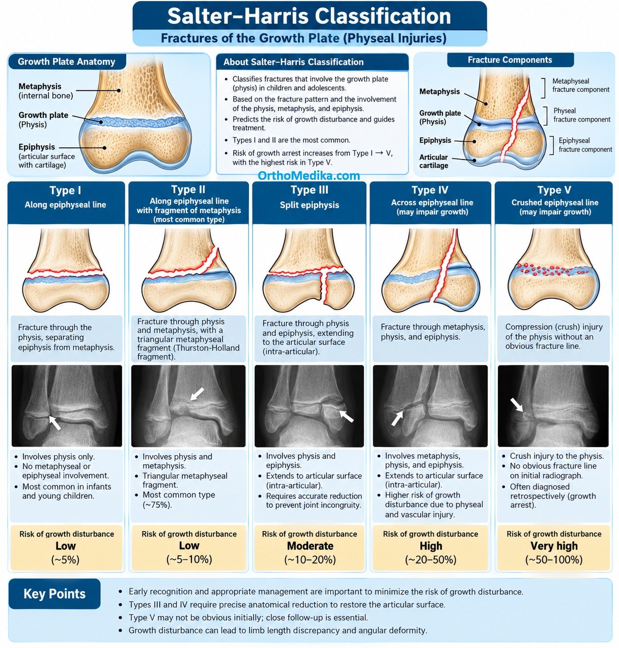
Salter–Harris fractures is that the growth plate is not one uniform layer. It is a stack of cartilage zones that continuously make new bone, and the fracture usually passes through the weakest layer while ideally sparing the “seed” cells needed for future growth.
How a long bone grows
A child’s long bone has two main ends:
- Epiphysis: the end of the bone, near the joint.
- Metaphysis: the wider part of the bone shaft next to the growth plate.
- Physis (growth plate): the cartilage layer between them; it makes the bone grow longer.
Think of the growth plate as a factory: new cartilage cells are produced near the epiphysis, mature as they move toward the metaphysis, then are replaced by bone. This process repeats and makes the bone longer.
Growth plate, layer by layer
Reading from the joint/epiphysis side toward the bone shaft/metaphysis side:
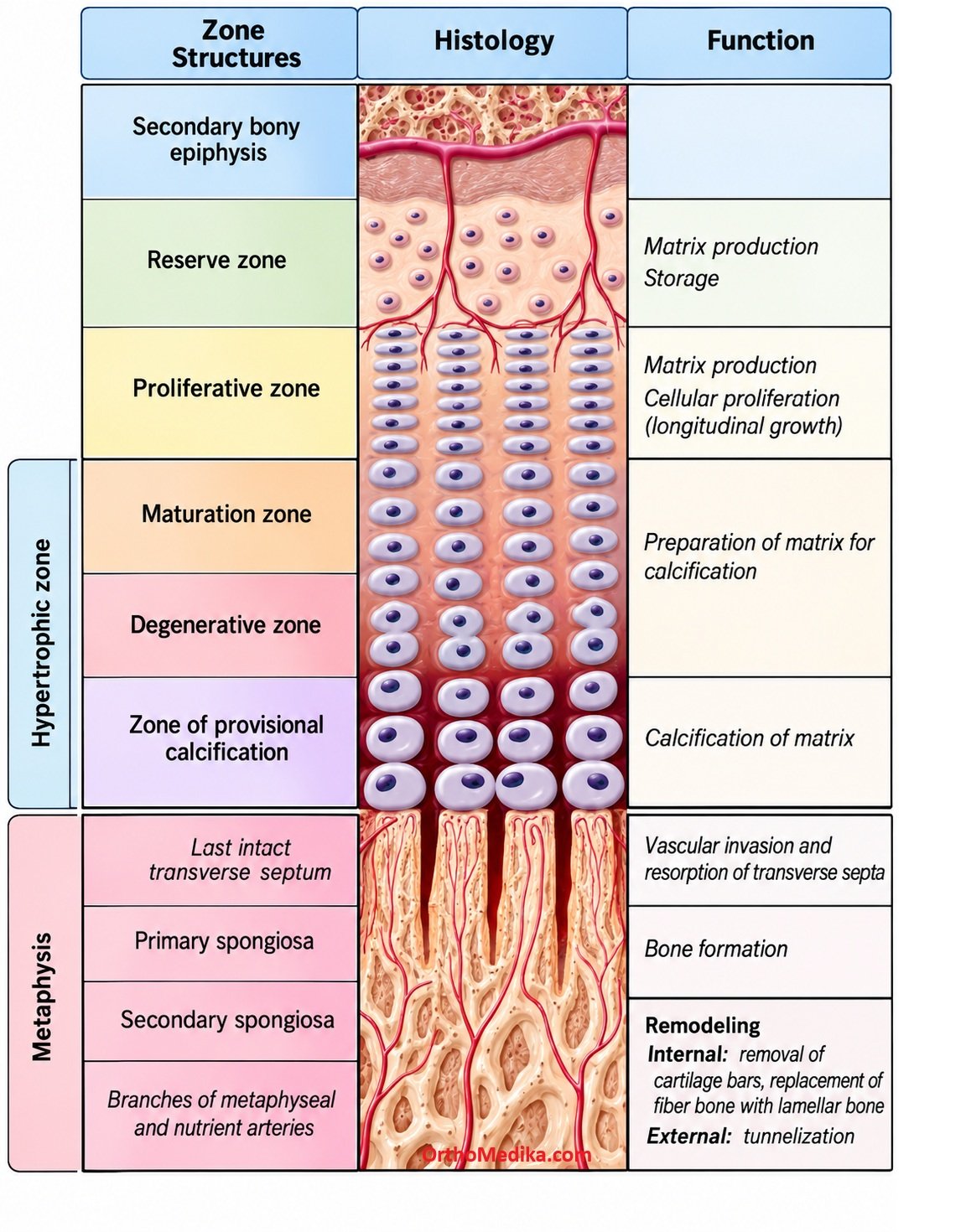
| Layer | Main job | What happens there |
| 1. Resting / reserve / germinal zone | Stores the “starter” or stem-like cartilage cells | Small, relatively inactive chondrocytes act as the source of future growth cells; this is the germinal layer. |
| 2. Proliferative zone | Makes more cartilage cells | Cells divide rapidly and arrange in columns parallel to the bone’s long axis; this is the major driver of longitudinal growth. |
| 3. Hypertrophic zone | Enlarges and matures cartilage cells | Chondrocytes become larger, mature, and eventually die; it includes maturation, degeneration, and early calcification stages. |
| 4. Zone of provisional calcification | Converts cartilage scaffold into bone | The cartilage matrix calcifies, blood vessels and bone-forming cells invade, and new bone is laid down on the metaphyseal side. |
A simple way to remember it: reserve cells → multiply → enlarge → calcify → become bone.
Why fractures occur in a specific layer
The hypertrophic zone, especially where it meets the calcified cartilage, is the weakest part of the growth plate. It has large cells and relatively less supporting matrix, so shear forces from a fall or twisting injury tend to split the plate there.
This is important because, in many Type I and II injuries, the fracture plane stays below the germinal and proliferative layers. The “seed cells” remain attached to the epiphysis and can continue functioning, which is why these injuries often have a good outlook.
How this explains Salter–Harris types
| Type | Fracture path | Effect on germinal/proliferative layers | Prognosis |
| I | Straight through the physis | Germinal cells usually remain with the epiphysis | Usually good |
| II | Through physis and a corner of metaphysis | Germinal cells usually remain intact | Usually good; most common type |
| III | Through physis and into the epiphysis/joint | Germinal/proliferative layers can be interrupted | Higher risk of growth disturbance, especially if displaced |
| IV | Crosses metaphysis, physis, and epiphysis | Directly disrupts the germinal/proliferative layers | High risk; often needs exact realignment or surgery |
| V | Crush/compression of the physis | Can damage multiple layers and blood supply | Rare but high risk of premature growth-plate closure |
Why the germinal layer matters
The germinal (resting/reserve) layer contains the cells that replenish the growth plate. If it is preserved and its blood supply remains intact, the plate can usually keep producing cartilage cells and the bone can continue growing normally.
If the germinal or proliferative layers are badly damaged—or if healing creates a bridge of bone across the plate—the growth plate may close early. This can cause:
- Limb shortening, if the whole plate stops growing.
- Angular deformity, if only one side of the plate stops growing while the other side continues.
- Joint problems, especially when the fracture extends into the epiphysis.
REFERENCES:
Levine RH, Thomas A, Nezwek TA, et al. Salter-Harris Fracture. [Updated 2023 Aug 10]. In: StatPearls [Internet]. Treasure Island (FL): StatPearls Publishing; 2023 Jan-. Available from: https://www.ncbi.nlm.nih.gov/books/NBK430688/
Waseem M, Taqi M, Marquart MJ. Pediatric Physeal Injuries Overview. [Updated 2024 Oct 1]. In: StatPearls [Internet]. Treasure Island (FL): StatPearls Publishing; 2024 Jan-. Available from: https://www.ncbi.nlm.nih.gov/books/NBK560546/